Univariate analysis on melody evaluation test
By Hsin-Yu Cheng
Published on June 12, 2025
June 12, 2025
Project definition
For brainhack school TW-SG 2025
bhfunc2025.py includes some functions are used in other code
projectVisualizeTest.ipynb is the main processing steps I work on extracting the activities and images from preprocessed fMRI images
The exported csv files are plotted to bar graphs in resultplotting.ipynb
Background
Inspired by the previous paper: Y.- A. Han and P.J. Hsieh Imaging Neuroscience, Volume 2, 2024 https://doi.org/10.1162/imag_a_00352. I want to understand how simple melody stimuli can induce neuronal activities in the brain.
Tools
The Univariate analysis on melody evaluation test project will rely on the following technologies:
- jupyter notebook
- python packages - nilearn and nibabel
- nilearn.dataset - Spatially constrained parcellation: msdl_rois
Data
Using the functional fMRI data in the previous paper: Y.- A. Han and P.J. Hsieh Imaging Neuroscience, Volume 2, 2024 https://doi.org/10.1162/imag_a_00352. Will not be provided when the project is uploaded.
Deliverables
- comparison of activity in a set of parcellated brain regions
Results
Progress overview
The preprocessed image series went through masking and activities are extracted.
Tools I learned during this project
- Github workflow
- dealing with Nifti data
- basic data visualization in seaborn
Results
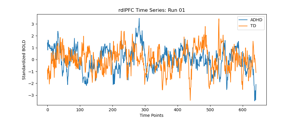
Deliverable 1: Comparison of activity difference
Two subjects, one run each, are analyzed to look for the activation difference in dorsal ACC, dlPFC, and a generalized auditory region.
Expected effect are seen in one run of one subject but the other doesn’t seem obvious.
Conclusion and acknowledgement
We can see the brain activity in different regions vary a lot in different individuals, even in identical test runs. More data needs analysis for a further, more general conclusion.